

Chemistry, Department of: Faculty Series
Robert Powers Publications
Accessibility Remediation
If you are unable to use this item in its current form due to accessibility barriers, you may request remediation through our remediation request form.
Document Type
Article
Date of this Version
1989
Citation
Published in Journal of Biomolecular Structure and Dynamics 7:3 (1989), pp. 515–556
Abstract
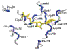
Assignment of the 1H and 31P NMR spectra of a decamer oligodeoxyribonucleotide duplex, d(CCCGATCGGG), and its quinoxaline ([MeCys3, MeCys7]TANDEM) drug duplex complex has been made by two-dimensional 1H-1H and heteronuclear 31P-1H correlated spectroscopy. The 31P chemical shifts of this 10 base pair oligonucleotide follow the general observation that the more internal the phosphate is located within the oligonucleotide sequence, the more upfield the 31P resonance occurs. While the 31P chemical shifts show sequence-specific variations, they also do not generally follow the Calladine “rules” previously demonstrated. 31P NMR also provides a convenient monitor of the phosphate ester backbone conformational changes upon binding of the drug to the duplex. Although the quinoxaline drug, [MeCys3, MeCys7]TANDEM, is generally expected to bind to duplex DNA by bis-intercalation, only small 31P chemical shift changes are observed upon binding the drug to duplex d(CCCGATCGGG). Additionally, only small perturbations in the 1H NMR and UV spectra are observed upon binding the drug to the decamer, although association of the drug stabilizes the duplex form relative to the other states. These results are consistent with a nonintercalative mode of association of the drug. Modeling and molecular mechanics energy minimization demonstrate that a novel structure in which the two quinoxaline rings of the drug binds in the minor groove of the duplex is possible.
Comments
Copyright © 1989 Adenine Press. Published by Taylor & Francis. Used by permission.