

Department of Physics and Astronomy: Individual Faculty Pages
Alexei Gruverman Publications
Accessibility Remediation
If you are unable to use this item in its current form due to accessibility barriers, you may request remediation through our remediation request form.
Document Type
Article
Date of this Version
11-10-2006
Abstract
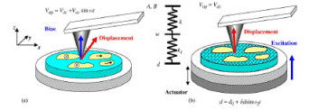
High-resolution studies of dental tissues are of considerable interest for biomedical engineering and clinical applications. In this paper, we demonstrate the application of piezoresponse force microscopy (PFM) to nanoscale imaging of internal structure of human teeth by monitoring the local mechanical response to an electrical bias applied via a conductive tip. It is shown that PFM is capable of detecting dissimilar components of dental tissues, namely, proteins and calcified matrix, which have resembling morphology but different piezoelectric properties. It is demonstrated that collagen fibrils revealed in chemically treated intertubular dentin exhibit high piezoelectric activity and can be visualized in PFM with spatial resolution of 10 nm. Evidence of the presence of protein inclusions of 100–200 nm wide and several micrometers long in tooth enamel has been obtained. Furthermore, it is found that the peritubular dentin and intertubular dentin exhibit different piezoelectric behavior suggesting different concentration of collagen fibrils. The obtained results demonstrate a high potential of PFM in providing an additional insight into the structure of dental tissues. It is suggested that the PFM approach can be used to study the structure of a wide range of biological materials by monitoring their electromechanical behavior at the nanoscale.
Comments
Published in Biochemical and Biophysical Research Communications 352 (2007) 142–146. Copyright © 2006 Elsevier Inc. Used by permission.