

Chemistry, Department of: Faculty Series
Robert Powers Publications
Accessibility Remediation
If you are unable to use this item in its current form due to accessibility barriers, you may request remediation through our remediation request form.
Document Type
Article
Date of this Version
1991
Citation
J. Mol. Biol. (1991) 221. 1081-1090
Abstract
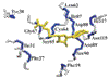
The solution structure of the ribonuclease H domain of HIV-l reverse transcriptase has been investigated by three-dimensional double and triple resonance heteronuclear magnetic resonance spectroscopy. The domain studied has 138 residues and comprises residues 427 to 560 of the 66 kDa reverse transcriptase with an additional four residues at the N terminus. Initial studies on the wild-type protein were hindered by severe differential line broadening, presumably due to conformational averaging. Mutation of the single tryptophan residue located in a loop at position 113 (position 535 in the reverse transcriptase sequence) to an alanine resulted in much improved spectral properties with no apparent change in structure. 1H, 15N and 13C backbone resonances were assigned sequentially using a range of three-dimensional double and triple resonance heteronuclear experiments on samples of uniformly (>95%) 15N and 15N/13C-labeled protein. and the secondary structure was elucidated from a qualitative analysis of data derived from three-dimensional 15N- and 13C-edited nuclear Overhauser enhancement spectra. The secondary structure comprises three α-helices and five strands arranged in a mixed parallel/antiparallel β-sheet with a + 1, + 1. -3x. -- 1 x topology. The C-terminal region from residue 114 onwards appears to be conformationally disordered in solution as evidenced by an almost complete absence of sequential and medium range nuclear Overhauser effects.
Comments
U.S. Government Work