Department of Physics and Astronomy: Publications and Other Research

Alexei Gruverman Publications
Document Type
Article
Date of this Version
January 2007
Abstract
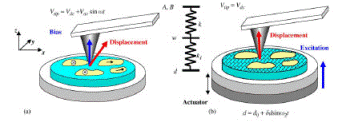
Hierarchical structure of connective and calcified tissues from the macro- to nanoscale level determines the mechanical and biological functionality of biological materials and has been the focus of numerous recent studies. Further progress in this field requires development of microscopic techniques capable of probing materials properties, including local composition, crystallographic orientation, and mechanical properties on the nanometer-length scale. Here, we describe a piezoresponse force microscopy (PFM) approach to high-resolution imaging of biological systems, based on detection of the local piezoelectric response. Samples include human tooth, femoral cartilage, deer antler, and butterfly wing scales. PFM allows differentiation between organic and mineral components and provides additional important information on materials microstructure. We also demonstrate the PFM capability of studying the internal structure and orientation of protein microfibrils with a spatial resolution of several nanometers. Future potential of the PFM approach for biological imaging is discussed.


Comments
Published in Scanning Probe Microscopy: Electrical and Electromechanical Phenomena at the Nanoscale, Sergei Kalinin and Alexei Gruverman, editors, 2 volumes (New York: Springer Science+Business Media, 2007). Used by permission.