

Plant Pathology, Department of
James Van Etten Publications
Accessibility Remediation
If you are unable to use this item in its current form due to accessibility barriers, you may request remediation through our remediation request form.
Document Type
Article
Date of this Version
2005
Citation
Microsc Microanal 11(Suppl 2), 2005, pp 1056-1057.
DOI: 10.1017/S1431927605500266
Abstract
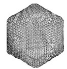
Paramecium bursaria chlorella virus 1 (PBCV-1) (genus Chlorovirus, family Phycodnaviridae), infects certain unicellular, exsymbiotic, chlorella-like green algae [1]. The PBCV-1 virion (~1x10⁹ Da) has a linear dsDNA genome (330kbp) and contains at least 100 different proteins [1, unpublished data]. A published cryo-electron microscopy and three-dimensional (3D) image reconstruction of PBCV-1 (26Å resolution) revealed that the outer icosahedral glycoprotein capsid, with a maximum diameter of 1,900Å along the five-fold axis, consists of 1680 trimeric capsomers (trimer) and 12 pentameric capsomers (pentamer) [2]. These capsomers are arranged on a T=169d (h=7, k=8) quasi-equivalent lattice. The trimers and pentamers are organized into 20 trisymmetrons (66 trimers) and 12 pentasymmetrons (30 trimers plus one central pentamer). In the present study, we have recorded new images, which have been used to compute a 3D image reconstruction of PBCV-1 at 15Å resolution. PBCV-1 sample preparation and microscopy were carried out as described [2]. Of the 94 micrographs recorded in an FEI/Philips CM200 FEG microscope (defocus level 1.44 ~ 3.64μm, x38,000), 45 were digitized at 7μm intervals on a Zeiss Phodis scanner. The obtained images were binned and interpolated to give a pixel size of 4.0Å. Particle origins and orientations were determined by model-based procedures with the 26Å map of PBCV-1 serving as the initial model [2, 3]. Corrections were made to each image to compensate for the effects of the microscope contrast transfer function. The effective resolution of the final reconstruction was determined by Fourier Shell Correlation analysis to be 15Å [4]. Of the 1262 boxed particle images, 1000 were used to compute the final density map. The latest reconstruction (Fig. 1A-D) reveals a more detailed view of the morphological features in the PBCV-1 virion compared to that observed in the 26Å map. The bilayer membrane is now more prominent and uniform. Also, each trimer, formerly seen as a simple, doughnut-like structure, now exhibits a hexagonal base that is topped with three, tower-like protrusions. The higher resolution PBCV-1 map permitted more accurate fitting of the atomic model for the Vp54 trimer into the virion density map and this has provided a more detailed view of the organization of the trisymmetron and pentasymmetron structures, and also may provide clues about the locations of minor capsid proteins [5]. One of the 28 trimers in each asymmetric unit is unique in that it contains a fiber-like structure (Fig. 1.B, black arrow) and these fibers (60/virion) may be involved in host recognition and attachment [6].
Comments
Copyright 2005 Microscopy Society of America