

Plant Pathology, Department of
James Van Etten Publications
Accessibility Remediation
If you are unable to use this item in its current form due to accessibility barriers, you may request remediation through our remediation request form.
Document Type
Article
Date of this Version
2018
Citation
Published (as Chapter 12) in Y. Yamaguchi, K. Kato (eds.), Glycobiophysics, Advances in Experimental Medicine and Biology 1104, pp 237-257.
doi 10.1007/978-981-13-2158-0_12
Abstract
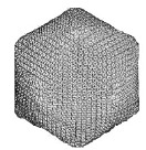
The capsid of Paramecium bursaria chlorella virus (PBCV-1) contains a heavily glycosylated major capsid protein, Vp54. The capsid protein contains four glycans, each N-linked to Asn. The glycan structures are unusual in many aspects: (1) they are attached by a β-glucose linkage, which is rare in nature; (2) they are highly branched and consist of 8–10 neutral monosaccharides; (3) all four glycoforms contain a dimethylated rhamnose as the capping residue of the main chain, a hyper-branched fucose residue and two rhamnose residues ''with opposite absolute configurations; (4) the four glycoforms differ by the nonstoichiometric presence of two monosaccharides, l-arabinose and d-mannose; (5) the N-glycans from all of the chloroviruses have a strictly conserved core structure; and (6) these glycans do not resemble any structures previously reported in the three domains of life.
The structures of these N-glycoforms remained elusive for years because initial attempts to solve their structures used tools developed for eukaryotic-like systems, which we now know are not suitable for this noncanonical glycosylation pattern. This chapter summarizes the methods used to solve the chlorovirus complex glycan structures with the hope that these methodologies can be used by scientists facing similar problems.
Comments
Copyright © 2018 Springer Nature Singapore Pte Ltd. Used by permission.